DOI: 10.59471/ijhsc202324
A specific reagent to study trypanosomatids respiration through polyprenyl diphosphate synthases inhibition: 1-[(n-oct-1-ylamino) ethyl] 1,1-bisphosphonic acid
Un reactivo reactivo para estudiar la respiración de tripanosomátidos mediante la inhibición de poliprenil difosfato sintasas: ácido 1-[(n-oct-1-ilamino) etil] 1,1-bisfosfónico
Octavio Fusco1 * ![]() , Cristina
Soraires2 *
, Cristina
Soraires2 * ![]() , Alicia
Hoffer1 *
, Alicia
Hoffer1 * ![]() , Alicia G.
Fuchs1,3 *
, Alicia G.
Fuchs1,3 * ![]() , Alina
Perrone1 *
, Alina
Perrone1 * ![]() , Patricia
Garavaglia1 *
, Patricia
Garavaglia1 * ![]() , Bruno
Travi4 *
, Bruno
Travi4 * ![]() , Juan
Carlos Engel5 *
, Juan
Carlos Engel5 * ![]() , Laurence
Lecordier6 *
, Laurence
Lecordier6 * ![]() , Laura
Fichera1 *
, Laura
Fichera1 * ![]() , Cristina
Maidana1 *
, Cristina
Maidana1 * ![]() , Victoria
Fragueiro Frías1 *
, Victoria
Fragueiro Frías1 * ![]() , Gabriela
A. García1 *
, Gabriela
A. García1 * ![]() , Jacqueline
Bua1,3 *
, Jacqueline
Bua1,3 * ![]() , Sergio H.
Szajnman7 *
, Sergio H.
Szajnman7 * ![]() , Juan
Bautista Rodríguez7 *
, Juan
Bautista Rodríguez7 * ![]() , Romina
Rebozzio1 *
, Romina
Rebozzio1 * ![]() , Mónica Esteva1
*
, Mónica Esteva1
* ![]() , Carlos
Pravia1 *
, Carlos
Pravia1 * ![]() , Benoît
Vanhollebeke6 *
, Benoît
Vanhollebeke6 * ![]() , Esteban
Bontempi Yela1 *
, Esteban
Bontempi Yela1 * ![]()
1Instituto Nacional de Parasitología “Dr. M. Fatala Chaben”, A.N.L.I.S/Malbrán, Secretaría de Salud. Buenos Aires 1063, Argentina.
2Pharmacology Department and IQUIMEFA (CONICET-FFYB), Facultad de Farmacia y Bioquímica, Universidad de Buenos Aires. 1113, Argentina.
3CAECIHS, Universidad Abierta Interamericana. Buenos Aires 1270, Argentina.
4Depts. of Internal Medicine & Microbiology and Immunology, University of Texas Medical Branch (UTMB). Galveston, Texas 77555-0435, USA.
5Sandler Center for Basic Research in Parasitic Diseases and Department of Pathology, University of California. San Francisco, California 94158-2330, USA.
6Laboratory of Molecular Parasitology, Institute of Molecular Biology and Medicine, Université Libre de Bruxelles. Gosselies B-6041, Belgium.
7Departamento de Química Orgánica and UMYMFOR (CONICET-FCEyN), Facultad de Ciencias Exactas y Naturales, Universidad de Buenos Aires. 1428, Argentina.
![]()
Submitted: 12-01-2023 Revised: 19-04-2023 Accepted: 15-07-2023 Published: 16-07-2023
How to Cite: Fusco O, Soraires C, Hoffer A, Fuchs AG, Perrone A, Garavaglia P, et al. A specific reagent to study trypanosomatids respiration through polyprenyl diphosphate synthases inhibition: 1-[(n-oct-1-ylamino) ethyl] 1,1-bisphosphonic acid. Interamerican Journal of Health Sciences. 2023; 3:24. https://doi.org/10.59471/ijhsc202324
ABSTRACT
Introduction: ubiquinone or Coenzyme Q (CoQ) is a molecule performing an essential function as an electron carrier in the respiratory chain. Besides, it functions as a proton and electron acceptor and as an antioxidant. It is composed of a benzoquinone ring and an isoprenoid chain, the last one being synthesized by a polyprenil diphosphate synthase. The inhibition of the Trypanosoma cruzi enzyme by 1-[(n-oct-1-ylamino) ethyl] 1,1-bisphosphonic acid (OBA) is lethal, as well as the interference of the homologous enzyme in T. brucei brucei. We now studied the OBA sensitivities of the different forms of human pathogenic trypanosomatids.
Methods: the different forms of trypanosomatids were cultured and their EC50 or LC50 were determined.
Results: we herein report that their mammalian forms (bloodstream forms of T. brucei, trypomastigotes and amastigotes of T. cruzi, as well as amastigotes of Leishmania donovani) are very sensitive to the compound (low micromolar half-maximal effective concentrations). Mammalian host cells were much less sensitive, and the Selectivity Indexes of the drug for the three trypanosomatids were established. Based on these results, we propose OBA as a useful reagent to study the respiratory pathway even in intracellular trypanosomatids.
KEYWORDS
Trypanosoma Cruzi, Trypanosoma Brucei, Leishmania, Solanesyl Diphosphate Synthase, Ubiquinone, Inhibitor, Bisphosphonate, EC50, LC50.
RESUMEN
Introducción: la ubiquinona o coenzima Q (CoQ) es una molécula que desempeña una función esencial como transportador de electrones en la cadena respiratoria. Además, funciona como aceptora de protones y electrones y como antioxidante. Está compuesta por un anillo de benzoquinona y una cadena isoprenoide, esta última sintetizada por una poliprenil difosfato sintasa. La inhibición de la enzima de Trypanosoma cruzi por el ácido 1-[(n-oct-1-ilamino) etil] 1,1-bisfosfónico (OBA) es letal, al igual que la interferencia de la enzima homóloga en T. brucei brucei. Ahora estudiamos la sensibilidad al OBA de las diferentes formas de tripanosomátidos patógenos humanos.
Método: se cultivaron las diferentes formas de tripanosomátidos y se determinó su EC50 o LC50.
Resultados: informamos de que sus formas en mamíferos (formas del torrente sanguíneo de T. brucei, tripomastigotes y amastigotes de T. cruzi, así como amastigotes de Leishmania donovani) son muy sensibles al compuesto (concentraciones efectivas semimáximas micromolares bajas). Las células hospedadoras de mamíferos fueron mucho menos sensibles, y se establecieron los índices de selectividad del fármaco para los tres tripanosomátidos. Basándonos en estos resultados, proponemos la OBA como un reactivo útil para estudiar la vía respiratoria incluso en tripanosomátidos intracelulares.
PALABRAS CLAVE
Trypanosoma Cruzi, Trypanosoma Brucei, Leishmania, Solanesil Difosfato Sintasa, Ubiquinona, Inhibidor, Bifosfonato, EC50, LC50.
INTRODUCTION
Ubiquinone or Coenzyme Q (CoQ) is a molecule that participates in essential metabolic functions, like respiration, redox equilibrium(1) and interaction with the Mitochondrial Permeability Transition Pore.(2,3) It is present in all cellular membranes.(4) Functionally, it is composed of a benzoquinone ring involved in redox cycling, and an isoprenoid tail allowing interaction with membranes.(5) In the yeast Saccharomyces cerevisiae, Coq is synthesized in a stepwise manner by different enzymes associated with the inner mitochondrial membrane.(6) The complex (Coq3-Coq9, and Coq11.(7) is called CoQ synthome (yeast) or Complex Q (human cells). The Polyprenyl diphosphate synthase (Coq1) is not included, but its product, the isoprenoid chain, is condensed to para-hydroxybenzoate by Coq 2 and then taken up by the complex (Coq3).
Polyprenyl diphosphate synthases share a common structural feature: the presence of a hydrophobic pocket where protruding amino acid residues determine the length of the chain that can be synthesized. Substrates bind to both aspartic rich motifs via an interaction with Mg2+.(8)
The isoprenoid tail has a specific size (or range of sizes) in each species. As nine isopentenyl units is the main size identified in Trypanosoma brucei and Trypanosoma cruzi CoQs(9,10) the enzyme is called Solanesyl Diphosphate Synthase (SPPS). We already described that in T. cruzi CoQ9 isoprenoid tail is synthesized by a glycosomal enzyme (TcSPPS)(11) for which a specific inhibitor, the 1-[(n-oct-1-ylamino)ethyl] 1,1-bisphosphonic acid (OBA), was designed.(12) The T. brucei homologue is a mitochondrial enzyme(13) whose knock down caused severe consequences very similar to those exerted by the inhibitor: depletion of the CoQ pool, reduced cell growth and oxygen consumption, and increased generation of reactive oxygen species.(14) In Leishmania there are no direct reports of SPPS presence, but CoQ with 7 to 10 isoprene units have experimentally been found in different stages of L. donovani, L. amazonensis and L. tarentolae.(15,16,17,18,19) This suggests the presence of several long chain (heptaprenyl, octaprenyl, solanesyl and decaprenyl) diphosphate synthases, or, alternatively, one enzyme producing different length products.(8)
Importantly, CoQ, the final product of this biosynthetic pathway, is present in the classical (terminal enzyme: cytochrome oxidase) respiratory chain as well as in the alternative respiratory chain (terminal enzyme: salicylhydroxamic acid -sensitive trypanosome alternative oxidase) present in Trypanosoma brucei. Because oxygen is the common final electron acceptor of these pathways, blocking CoQ synthesis would be similar to subjecting the parasite to a combined treatment with cyanide and salicylated hydroxamic acid (inhibiting both terminal oxidases), or to anaerobiosis.
The metabolic strategies that have been described in the different trypanosomatid parasites under anaerobic conditions are the following:
T. brucei –Procyclic and bloodstream forms are extracellular and depend on the mitochondrial respiratory chains to reoxidize the reduction equivalents generated by the glucose and amino acids metabolisms. In anaerobiosis the insect form (procyclics) can neither reoxidize the glycosomal NADH through the alternative oxidase (TAO) nor the mitochondrial NADH through the classical chain. To reach glycosomal and mitochondrial equilibrium they could secrete reduced molecules such as acetate, succinate, lactate and alanine.(21)
The mammalian form (bloodstreams) is primarily glucose-dependent and has reduced mitochondrial functions. The main end product in aerobiosis is pyruvate, and the redox equilibrium is reached through the glycerol 3-phosphate/dihydoxyacetone phosphate shuttle and the mitochondrial TAO. As this last step is sensitive to oxygen deprivation, in anaerobiosis the parasite reoxidize NADH by converting dihydroxyacetone phosphate to glycerol, which is excreted at equal amounts as pyruvate.(21,22,23) Following this pathway the parasites resign the production of one ATP per triose phosphate.(20) The anaerobiosis result is coincident with the TAO mRNA interference experiment:(24) induction of interference almost doubled the duplication time, and diminished three quarters the oxygen consumption.
T. cruzi – The parasite adopt mainly three forms: extracellular in insects (epimastigotes) and in mammals (trypomastigotes), and intracellular in mammals (amastigotes). In the first two, the final (secreted) products of glucose metabolism in aerobiosis are succinate, L-alanine and acetate, while in anaerobiosis there is an increased secretion of succinate. Trypomastigotes also produced L-lactate in anaerobiosis.(23,25)
Amastigotes were considered to rely on amino and fatty acids. But recent experiments showed that radiolabelled glucose was incorporated by isolated amastigotes into glycolytic and Krebs cycle intermediates. Moreover, addition of exogenous glucose increased their oxygen consumption rate and extracellular acidification rate, while glucose restriction elongated by 50 % the division time.(26) Thus, this form of the parasite could also be sensitive to anaerobic conditions.
Leishmania - Promastigotes (extracellular insect form) secreted also several reduced molecules in anaerobic conditions: succinate, acetate, pyruvate, glycerol and D-lactate produced through the methylglyoxal pathway.(27,28) Upon differentiation, amastigotes (intracellular mammalian forms) enter a semi-quiescent growth state, consuming less glucose and amino acids. This nutrient-sparing metabolism was called the stringent metabolic response.(29) The main intracellular glucose catabolites in anaerobically grown axenic L. donovani and L. pifanoi amastigotes were alanine and acetate, while the secreted products contained also succinate and glycerol.(30)
The glucose catabolism is essential in Leishmania species, but curiously gluconeogenesis is as well(31) highlighting the importance of the anabolic enzyme fructose-1,6-bisphosphatase in the synthesis of glycoconjugates and nucleic acids.(32) The acetyl-CoA and end-products from the glucose catabolism produce glutamate, which is involved in thiols, pyrimidine and amino sugar synthesis. The glutamate production step (inhibited by sodium fluoroacetate) seems to be essential, as depletion of the glutamate intracellular pool can not be rescued by exogenous uptake.(29) The amino acid catabolism and fatty acid β-oxidation are up-regulated.(33) Several of the mentioned pathways produce reducing equivalents that have to be reoxidized through the respiratory chain, otherwise their accumulation could stop the whole metabolism. Thus, it is of interest to determine CoQ involvement in these processes.
In trypanosomatids SPPS synthesizes the isoprenoid moiety of CoQ, molecule centrally involved in both respiratory chains. It is of interest to determine the OBA sensitivity of the remaining trypanosomatids and forms, to postulate this molecule as an useful reagent to study trypanosomatids respiration.
METHOD
Extracellular forms. Determination of EC50 and LC50
Parasites were adjusted to an initial concentration of 5 x 10⁴ mL-1 (T. brucei bloodstream form, T. cruzi epimastigotes, Leishmania donovani and L. major promastigotes), 5 x 10⁵ mL-1 (L. braziliensis and L. m. amazonensis promastigotes) or 1 x 10⁶ mL-1 (T. brucei procyclic form) in 200 µL medium and loaded into sterile 96-well plates. Two-fold serial dilutions of OBA, boiled to ensure complete dissolution and sterility, were added to duplicate wells. After 3 days incubation (37°C-5 % CO2, or 28°C), cells in all wells were counted by using a Neubauer hemocytometer or a Beckman Coulter Counter. Each assay was repeated at least three times. The EC50 (effective concentration for half-maximal growth inhibition) was determined by linear and polynomial regression using the Origin software version 6 (OriginLab Corporation, USA).
T. cruzi bloodstream trypomastigotes (Tulahuen strain, Tul 2 stock) were obtained by cardiac puncture from infected BALB/c mice. Trypomastigotes (5 x 10⁵ cells mL-1) were incubated with different drug dilutions in polystyrene 96-well U-plates at 4°C during 24 h. Each well was then microscopically examined under an 18 mm diameter coverslip at 400 magnification. The percentage of lysis was calculated considering the untreated control as 100 %. The LC50 (half-maximal lethal concentration) was defined as the minimal concentration of compound required to lyse 50 % of the trypomastigotes.
Bloodstream forms of T. b. rhodesiense Etat 1,2R (1 x 10³ cells mL-1) and T. b. gambiense LiTat 1,3 (4 x 10³ cells mL-1) were loaded in triplicates in white 96 well plates (Lumitrac) with serial dilutions of the drug in a total volume of 50 µL. After 72h incubation at 37°C-5 % CO2, cell density was estimated comparing the ATP level in treated versus untreated wells (assuming a constant ATP amount per cell, see comments in Discussion) (CellTiter-Glo, Promega).
Intracellular forms. Determination of EC50
For Leishmania amastigotes we used an Ex Vivo Splenic Explant Model System.(34) Two hamsters were infected with a Leishmania strain transfected with an episomal vector containing the luciferase (luc) reporter gene.(35) Splenocytes were purified, counted, and 100 000 added to each well of a 96-well sterile white-bottom plate (Costar) in 50 µL of culture medium. Subsequently, the same volume of different OBA concentrations were added to each well and the microplate was incubated for 48 hours at 37°C in a humidified atmosphere and 5 % CO2. Finally, cells were lysed and the luciferase activity determined in a plate luminometer following the addition of 100 µL of luciferin substrate at room temperature (Promega). The luminometry counts were transformed to number of parasites using a linear standard curve of luciferase activity versus number of microscopically enumerated amastigotes.
For T. cruzi amastigotes we used a high-throughput assay based on automatic image acquisition and analysis.(36) Briefly, host mammalian cells (bovine embryo skeletal muscle BESM and human hepatoma Huh-7) grown on 96-well plates were infected with CA-I/72 trypomastigotes. Sixteen hours later different dilutions of OBA were added and cells were further incubated for 72 h. Plates were washed, fixed and then stained with the DNA fluorescent dye DAPI (4’,6-diamidino-2-phenylindole dihydrochloride). Analysis of the acquired images (IN Cell Analyzer 1000, GE Healthcare) allowed detecting the number of cell nuclei and parasite kinetoplasts. Data analysis produced not only the number of amastigotes per cell, but also the density of host cells per field (toxicity).
Cytotoxicity assay
Vero cells were grown in RPMI medium, supplemented with 10 % FBS, 100 IU/mL penicillin and 0,1 mg/mL streptomycin, at 37°C in the presence of 5 % CO2. Cells were seeded in 96-well plates at a density of 1 x 10⁴ cells/ well and left untouched for 24 h. After incubation, the growing cells were treated with different concentrations of OBA (10-500 µM) or DMSO (negative controls) for an additional 72 h. Subsequently, the culture medium was removed and 3-(4,5-dimethylthiazol-2yl)-2,5-diphenyltetrazolium bromide (MTT) from a stock solution (5mg/mL) was added at a final concentration of 0,75mg/mL. After a 2 h incubation, the blue precipitates were dissolved in 100 µL of dimethyl sulfoxide (DMSO) and were read on a plate reader (Mindray MR-96A) at a wavelength of 578 nm with a reference filter of 630 nm. Each test was performed in triplicate. The 50 % cytotoxic concentration (CC50) was determined using the GraphPadPrism 6,01 software.
HepG2 cells (human hepatocellular carcinoma, ATCC#HB 8065) were grown and cytotoxicity evaluated as described.(34)
RESULTS
In table 1 we summarize our current and previously published results characterizing OBA sensitivity of each trypanosomatids life stage.
T. brucei has no intracellular forms, being present in tsetse flies as procyclic, epimastigote and metacyclic forms, and in mammals (the bloodstream stage) only as slender and stumpy forms. We have already reported that the EC50 for T. b. brucei, the species responsible for the animal disease nagana, was 50 µM and 2 µM, for procyclics and bloodstream parasites respectively.(14) We now determined that the EC50 for the mammalian form of T. b. rhodesiense and T. b. gambiense was even lower (0,92 µM and 0,78 µM, respectively).
The other parasitic species included in this study (Leishmania spp. and Trypanosoma cruzi) have extracellular and intracellular forms. To assess the sensitivity of T. cruzi epimastigotes to OBA, we tested CL Brener clone(37) and Tc Nicaragua isolate(38) belonging to different Discrete Typing Units (DTU VI and DTU I, respectively). We used drug concentrations ranging from 50 to 800 µM. Both EC50s were very close (270 and 209 µM), suggesting that both strains share similar mechanisms of drug uptake, modification and excretion.
Regarding the intracellular stage, we reported for Y strain amastigotes growing within L6E9 myoblasts an EC50 of 4,1 µM.(12) To confirm that result we used an image-based technique(36) able to distinguish between cell nuclei and amastigote kinetoplasts. This method yielded an EC50 of 3,13 µM for clone CA-I/72.
Since trypomastigotes sensitivity to OBA cannot be evaluated by growth differences, as it is a non-replicative stage, we measured lysis of drug-treated parasites. As can be observed in Table 1, the LC50 value was intermediate between the ones obtained with epimastigotes and amastigotes.
In the case of Leishmania we first searched for the target of OBA. In tritrypdb(39) only one gene with similarity to TcSPPS was detected in the different species (L. donovani, L. major, L. braziliensis, L. mexicana, L. infantum, L. tropica, L. aethiopica, etc). The retrieved protein sequences were aligned using Clustal Omega.(40) They were very similar to TcSPPS (more than 75 % homology), and exhibited the seven conserved domains for enzymatic activity(41) (figure 1). The two essential aspartic rich motifs, located in domains II and VI, and the small residues before the first motif (AS) allow its classification as a long chain polyprenyl diphosphate synthase. These enzymes synthesize the isoprenic chain that is then attached to the benzoquinone ring of CoQ. The EC50 evaluations carried out using cultured promastigotes showed that only L. mexicana amazonensis was sensitive to OBA while L. braziliensis, L. major, and L. donovani were significantly more resistant.
To test the intracellular stage of Leishmania parasites we used a recently developed ex vivo model able to measure amastigote multiplication in macrophages under the potential influence of lymphocytes within the splenic environment.(34) L. major (cutaneous leishmaniasis) amastigotes were not affected by the treatment with the highest concentration of OBA (10 µM during 48 hs). On the other hand, L. donovani (visceral leishmaniasis) amastigotes were more sensitive to OBA, with an EC50 of 3,3 µM.
Finally, OBA toxicity was evaluated. The host cell growth in the high-throughput assay (bovine embryo skeletal muscle and human hepatoma Huh-7 cells) was not affected up to the highest tested concentration (12,5 µM). However, we included additional OBA cytotoxicity evaluation using Vero and HepG2 cells. We found that only high concentrations of the drug showed signs of toxicity in these cells (CC50, half-maximal cytotoxic concentration, 185,9 µM and 105,8 µM, respectively). This allowed to calculate the selectivity indexes of the compound (CC50 / EC50) for the mammalian forms of the parasites (table 1). Bloodstream african trypanosomes showed the higher selectivity index, followed by the intracellular stages.
DISCUSSION
Bisphosphonates have been studied as antiparasitic agents, targeted mainly to the essential enzyme farnesyl pyrophosphate synthase.(42,53) In this paper, we used an octyl containing bisphosphonate inhibiting specifically the enzyme SPPS. We will now discuss in detail the OBA sensitivity of several forms of the different trypanosomatids.
T. brucei procyclics secrete reduced molecules. This mechanism could explain the parasite resistance to OBA (EC50: 50 µM).
The mammalian (bloodstream) forms are more sensitive: 2 µM for T. b. brucei, and less for the two subspecies responsible for sleeping sickness in humans. It should be noted that the numbers for T. b. rhodesiense and T. b. gambiense should be slightly corrected. The indirect parasite quantification method was based on the ATP level, and we already reported a 50 % reduction of ATP content upon a 2 µM OBA treatment.(14) The corrected numbers are still well separated from the procyclics value, implying essential metabolic differences between them.
Another detail is that in respiration deficient conditions, the glycerol excreted by the bloodstream parasites and accumulating in the well plates could affect parasite metabolism. Within certain limits, glycerol shows no toxicity. For example, through the course of infection, bloodstream parasites are subjected and can metabolize low (intravascular, microMolar) and high (extravascular, miniMolar) glycerol concentrations.(44) But in other conditions, glycerol affects viability. Glycerol kinase inhibition by mass action (4 mM glycerol) rendered the parasite more susceptible to ascofuranone, a TAO inhibitor(45) and bloodstream forms adapted to grow in glycerol-rich medium (10 mM) are more sensitive to TAO inhibition.(46) In our published results (iRNA of the TbSPPS gene), the glycerol effect was minimized as cells were diluted to 105 cells ml-1 in fresh medium every other day, incidentally eliminating the excreted glycerol. (14) To further reject the cytotoxic effect of glycerol, we repeated the OBA sensitivity determination diluting the parasites back to the original concentration (35 x 103 ml-1) every day. We observed that the growth inhibitory effect of OBA persists at the same level, being independent of the accumulated glycerol.
The South American parasite T. cruzi also showed stage dependendent OBA sensitivity. Epimastigotes were resistant (200 µM), probably secreting reduced molecules to survive.
Because T. cruzi strains are grouped into 7 different DTUs(47) and we found fixed mutations in the SPPS gene that could affect the enzymatic behaviour (unpublished results), epimastigotes from two different strains were studied. No significant differences were found in terms of OBA sensitivity.
T. cruzi trypomastigotes were more sensitive to OBA (25 µM). Incidentally, the lysis assay allowed to confirm that, at least in this stage, OBA is not trypanostatic but trypanocidal.
T. cruzi amastigotes, the intracellular forms, were the most sensitive ones (4 µM). The EC50 value for clone CA-I/72 is very close to the previously reported for the Y strain.
In Leishmania the “overflow” metabolism could protect promastigotes from CoQ absence. The word refers to the secretion of reduced molecules taking place in high glucose or low oxygen concentrations.(48)
Leishmania amastigote sensitivity, on the other hand, is more difficult to predict. In principle, they should be more resistant to CoQ shortage due to the stringent response.(32,49) It is characterized by a slow growth and a metabolic reconfiguration. Specifically, as the TCA cycle is switched to anabolic tasks, the parasite should not be affected by OBA. Moreover, inside the phagolysosome it is immersed into a harsh environment (pH 4,5–5,5). Although the pH gradient is beneficial for substrate acquisition(50) it has to maintain the intracellular neutral pH by ATPases with energy expenditure.(51) ATP could be generated by substrate-level synthesis, which is also OBA independent. Thus, it is interesting that L. donovani amastigotes were very sensitive to OBA. The difference with L. major amastigotes does not seem to reside in the number of cells sharing every vesicle, as both Old World species dwell in single vacuoles.(52)
To account for the OBA sensitivity of some trypanosomatids intracellular forms, there is still an alternative explanation. Bisphosphonates show high affinity for calcium, sequestering it from solution. Some organisms, trypanosomatids included, possess a specialized calcium and phosphorus compounds-rich organelle called acidocalcisome.(53) It has been reported a higher concentration of these organelles (representing a higher percentage of the total cell volume) in amastigotes of T. cruzi and Leishmania major, in comparison with the other stages.(54,55) A possible hypothesis is that in contact with the inhibitor, these organelles could store some OBA, while the remnant soluble molecules could inhibit SPPS and begin to deplete the ATP pool. This could impair the action of the proton pumps that maintain the acidic pH of the organelles.(53) In alcaline conditions pyrophosphates will be hydrolyzed, releasing the chelated calcium and OBA, with lethal consequences. In this way, acidocalcisomes could function as controlled-release devices, whose action is more evident in acidocalcisome-rich stages.
In summary, the higher drug sensitivity of the intracellular stages compared with the axenically grown parasites, is puzzling, highlighting the importance of factors so far unknown. These unexplored features, including the pathway followed by the drug to traverse several cellular membranes and to reach a lethal level inside the intracellular amastigotes, deserve further research.
Finally, we determined OBA cytotoxicity and selectivity indexes. As can be seen in table 1, OBA is much more specific to parasitic enzymes than to mammalian ones. As a conclusion, this molecule could be a useful reagent to discriminate between trypanosomatids and host cells respiratory metabolisms.
|
Table 1. Sensitivity of the different forms of trypanosomatids to OBA |
||||
|
Cell |
Strain |
Form |
EC50a (µM) |
Selectivity Index |
|
T. b. brucei |
427 29-13 |
procyclic |
50 * |
- |
|
T. b. brucei |
SM, 427 90-13 |
bloodstream |
2 *, 1,07 ± 0,02 |
73 |
|
T. b. rhodesiense |
ETat 1,2R |
bloodstream |
0,92 ± 0,06 |
158,5 |
|
T. b. gambiense |
LiTat 1,3 |
bloodstream |
0,78 ± 0,01 |
187 |
|
T. cruzi |
Brener |
epimastigote |
270 ± 47 |
- |
|
Tc Nicaragua |
epimastigote |
209,6 ± 44 |
- |
|
|
Tulahuen, stock Tul 2 |
trypomastigote |
25 ± 2,3 ** |
5,8 |
|
|
Y |
amastigote |
4,1 *** |
35,5 |
|
|
CA-I/72 |
amastigote |
3,13 ± 0,24 |
46,6 |
|
|
L. braziliensis |
MHOM/BR/75/M2903 |
promastigote |
63,5 ± 12,3 |
- |
|
L. m. amazonensis |
IFLA/BR/1967/PH 8 |
promastigote |
4 ± 0,3 |
- |
|
L. major |
MHOM/IL/81/Friedlin |
promastigote |
> 200 |
- |
|
L. donovani |
MHOM/SD/001S-2D |
promastigote |
192 ± 7,2 |
- |
|
L. major |
MHOM/IL/81/Friedlin |
amastigote |
>10 |
ND |
|
L. donovani |
MHOM/SD/001S-2D |
amastigote |
3,3 ± 0,4 |
44,2 |
|
aEC50, half-maximal effective concentration, *(14), **LC50, half-maximal lethal concentration, ***(12), ND: not determined |
||||
Gene accession numbers (tritrypdb): LBRM2903_150017100, LbrM.15,2.001060, LmxM.15,1020, LdBPK_151080,1, LINF_150018400, LmjF.15,1020, LMJLV39_150017200, LMJSD75_150017200, LTRL590_150016700, LAEL147_000219000.

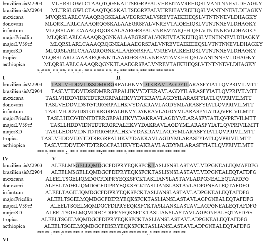
Figure 1. Comparison at the protein level of the long chain polyprenyl diphosphate synthase in different Leishmania species. (*) identical residue, (:) strong conservation, (.) weak conservation. Essential domains are highlighted and numbered.
Abbreviations
CoQ: Coenzyme Q; OBA: 1-[(n-oct-1-ylamino) ethyl] 1,1-bisphosphonic acid; SPPS: Solanesyl Diphosphate Synthase; EC50: effective concentration for half-maximal growth inhibition; LC50: half-maximal lethal concentration; CC50: drug concentration that reduces the cell viability by 50 %; TAO: trypanosome alternative oxidase.
REFERENCES
1. Wang Y, Hekimi S. Molecular genetics of ubiquinone biosynthesis in animals. Crit Rev Biochem Mol Biol. 2013;48(1):69-88.
2. Fontaine E, Ichas F, Bernardi P. A ubiquinone-binding site regulates the mitochondrial permeability transition pore. J Biol Chem. 1998;273:25734–40.
3. Bustos PL, Perrone AE, Milduberger NA, Bua J. Mitochondrial permeability transition in protozoan parasites: what we learned from Trypanosoma cruzi. Cell Death Dis. 2017 Sep 21;8(9):e3057.
4. Kawamukai M. Biosynthesis and applications of prenylquinones. Biosci Biotechnol Biochem. 2018;82(6):963-77.
5. Crane FL. Biochemical functions of coenzyme Q10. J Am Coll Nutr. 2001;20:591–8.
6. Kawamukai M. Biosynthesis of coenzyme Q in eukaryotes. Biosci Biotechnol Biochem. 2016;80(1):23-33.
7. Allan CM, Awad AM, Johnson JS, Shirasaki DI, Wang C, Blaby-Haas CE, et al. Identification of Coq11, a new coenzyme Q biosynthetic protein in the CoQ-synthome in Saccharomyces cerevisiae. J Biol Chem. 2015;290(12):7517-34.
8. Wang KC, Ohnuma S. Isoprenyl diphosphate synthases. Biochim Biophys Acta 2000;1529 (1-3):33-48.
9. Clarkson AB, Bienen EJ, Pollakis G, Grady RW. Respiration of bloodstream forms of the parasite Trypanosoma brucei brucei is dependent on a plant-like alternative oxidase. J Biol Chem. 1989;264:17770–6.
10. Löw P, Dallner G, Mayor S, Cohen S, Chait BT, Menon AK. The mevalonate pathway in the bloodstream form of Trypanosoma brucei. Identification of dolichols containing 11 and 12 isoprene residues. J Biol Chem. 1991;266(29):19250-7.
11. Ferella M, Montalvetti A, Rohloff P, Miranda K, Fang J, Reina S, et al. A solanesyl-diphosphate synthase localizes in glycosomes of Trypanosoma cruzi. J Biol Chem 2006;281:39339-48.
12. Szajnman SH, García Liñares GE, Li ZH, Jiang C, Galizzi M, Bontempi EJ, et al. Synthesis and biological evaluation of 2-alkylaminoethyl-1,1-bisphosphonic acids against Trypanosoma cruzi and Toxoplasma gondii targeting farnesyl diphosphate synthase. Bioorg Med Chem 2008;16:3283–90.
13. Lai DH, Bontempi EJ, Lukeš J. Trypanosoma brucei solanesyl-diphosphate synthase localizes to the mitochondrion. Mol Biochem Parasitol. 2012;183(2):189-92.
14. Lai DH, Poropat E, Pravia C, Landoni M, Couto AS, Rojo FG, et al. Solanesyl diphosphate synthase, an enzyme of the ubiquinone synthetic pathway, is required throughout the life cycle of Trypanosoma brucei. Euk Cell 2014;13(2):320-8.
15. Biswas S, Barrett MP, Rivière L, Bringaud F. Participation of chlorobiumquinone in the transplasma membrane electron transport system of Leishmania donovani promastigote: effect of near-ultraviolet light on the redox reaction of plasma membrane. Bioch Bioph Acta. 2008;1780(2):116-27.
16. Rassam MB, Shanshal M, Gargees GS. Isolation and identification of coenzyme Q from Leishmania donovani. Mol Biochem Parasitol. 1988;29(1):61-4.
17. Ellis JE, Setchell KD, Kaneshiro ES. Detection of ubiquinone in parasitic and free-living protozoa, including species devoid of mitochondria. Mol Biochem Parasitol. 1994;65(2):213-24.
18. Arruda DC, D’Alexandri FL, Katzin AM, Uliana SR. Leishmania amazonensis: biosynthesis of polyprenols of 9 isoprene units by amastigotes. Exp Parasitol 2008;118(4):624-8.
19. Ranganathan G, Mukkada AJ. Ubiquinone biosynthesis in Leishmania major promastigotes. Int J Parasitol. 1995;25(3):279-84.
20. Besteiro S, Barrett MP, Rivière L, Bringaud F. Energy generation in insect stages of Trypanosoma brucei: metabolism in flux. Trends Parasitol. 2005;21(4):185-91.
21. Grant PT, Fulton JD. The catabolism of glucose by strains of Trypanosoma rhodesiense. Biochem J. 1957;66:242-50.
22. Brohn FH, Clarkson AB. Jr. Quantitative effects of salycylhydroxamic acid and glycerol on Trypanosoma brucei glycolysis in vitro and in vivo. Acta Trop. 1978;35:23-33.
23. Ryley JF. Studies on the metabolism of the Protozoa. 7. Comparative carbohydrate metabolism of eleven species of trypanosome. Biochem J. 1956;62(2):215-22.
24. Helfert S, Estévez AM, Bakker B, Michels P, Clayton C. Roles of triosephosphate isomerase and aerobic metabolism in Trypanosoma brucei. Biochem J. 2001;357:117-25.
25. Cazzulo JJ, Arauzo S, Franke de Cazzulo BM, Cannata JJ. On the production of glycerol and L-alanine during the aerobic fermentation of glucose by trypanosomatids. FEMS Microbiol Lett. 1988;51:187-92.
26. Shah-Simpson S, Lentini G, Dumoulin PC, Burleigh BA. Modulation of host central carbon metabolism and in situ glucose uptake by intracellular Trypanosoma cruzi amastigotes. PLoS Pathog. 2017;13(11):e1006747.
27. Darling TN, Balber AE, Blum JJ. A comparative study of D-lactate, L-lactate and glycerol formation by four species of Leishmania and by Trypanosoma lewisi and Trypanosoma brucei gambiense. Mol Biochem Parasitol. 1988;30(3):253-7.
28. Irsch T, Krauth-Siegel RL. Glyoxalase II of African trypanosomes is trypanothione-dependent. J Biol Chem. 2004;279(21):22209-17.
29. Saunders EC, Ng WW, Kloehn J, Chambers JM, Ng M, McConville MJ. Induction of a stringent metabolic response in intracellular stages of Leishmania mexicana leads to increased dependence on mitochondrial metabolism. PLoS Pathog. 2014;10(1):e1003888.
30. Rainey PM, MacKenzie NE. A carbon-13 nuclear magnetic resonance analysis of the products of glucose metabolism in Leishmania pifanoi amastigotes and promastigotes. Mol Biochem Parasitol. 1991;45(2):307-15.
31. Naderer T, Ellis MA, Sernee MF, De Souza DP, Curtis J, Handman E, et al. Virulence of Leishmania major in macrophages and mice requires the gluconeogenic enzyme fructose-1,6-bisphosphatase. Proc Natl Acad Sci USA. 2006;103:5502-7.
32. McConville MJ, Saunders EC, Kloehn J, Dagley MJ. Leishmania carbon metabolism in the macrophage phagolysosome- feast or famine? F1000Res. 2015;4(F1000 Faculty Rev):938.
33. Rosenzweig D, Smith D, Opperdoes F, Stern S, Olafson RW, Zilberstein D. Retooling Leishmania metabolism: from sand fly gut to human macrophage. FASEB J. 2008;22:590-602.
34. Osorio Y, Travi BL, Renslo AR, Peniche AG, Melby PC. Identification of Small Molecule Lead Compounds for Visceral Leishmaniasis Using a Novel ExVivo Splenic Explant Model System. PLoS Negl Trop Dis. 2011;5(2):e962.
35. Roy G, Dumas C, Sereno D, Wu Y, Singh AK, Tremblay MJ, et al. Episomal and stable expression of the luciferase reporter gene for quantifying Leishmania spp. infections in macrophages and in animal models. Mol Biochem Parasitol 2000;110:195–206.
36. Engel JC, Ang KK, Chen S, Arkin MR, McKerrow JH, Doyle PS. Image-based high-throughput drug screening targeting the intracellular stage of Trypanosoma cruzi, the agent of Chagas’ disease. Antimicrob Agents Chemother. 2010;54(8):3326-34.
37. Zingales B, Pereira ME, Oliveira RP, Almeida KA, Umezawa ES, Souto RP, et al. Trypanosoma cruzi genome project: biological characteristics and molecular typing of clone CL Brener. Acta Tropica. 1997;68:159–73.
38. Grosso NL, Búa J, Perrone AE, Gonzalez MN, Bustos PL, Postan M, et al. Trypanosoma cruzi: biological characterization of a isolate from an endemic area and its susceptibility to conventional drugs. Exp Parasitol. 2010;126(2):239-44.
39. Aslett M, Aurrecoechea C, Berriman M, Brestelli J, Brunk BP, Carrington M, et al. TriTrypDB: a functional genomic resource for the Trypanosomatidae. Nucleic Acids Res. 2010;38 (Release 40. 15 Oct 2018):D457-62.
40. Madeira F, Park YM, Lee J, Buso N, Gur T, Madhusoodanan N, et al. The EMBL-EBI search and sequence analysis tools APIs in 2019. Nucleic Acids Res. 2019;47(W1):W636-41.
41. Koyama T. Molecular analysis of prenyl chain elongating enzymes. Biosci Biotechnol Biochem. 1999;63:1671–6.
42. Rodríguez JB, Falcone BN, Szajnman SH. Approaches for Designing new Potent Inhibitors of Farnesyl Pyrophosphate Synthase. Expert Opin Drug Discov. 2016;11(3):307-20.
43. Wolf K, Dormeyer M. Information-based methods in the development of antiparasitic drugs. Parasitol Res. 2003;90 Suppl 2:S91-6.
44. Bringaud F, Plazolles N, Pineda E, Asencio C, Villafraz O, Millerioux Y, et al. (2021) Glycerol, a possible new player in the biology of trypanosomes. PLoS Pathog 17(12): e1010035.
45. Minagawa N, Yabu Y, Kita K, Nagai K, Ohta N, Meguro K, et al. An antibiotic, ascofuranone, specifically inhibits respiration and in vitro growth of long slender bloodstream forms of Trypanosoma brucei brucei. Mol Biochem Parasitol. 1997;84:271-80.
46. Pineda E, Thonnus M, Mazet M, Mourier A, Cahoreau E, Kulyk H, et al. Glycerol supports growth of the Trypanosoma brucei bloodstream forms in the absence of glucose: Analysis of metabolic adaptations on glycerol-rich conditions. PLoS Pathog. 2018;14(11):e1007412.
47. Zingales, B., 2018. Trypanosoma cruzi genetic diversity: something new for something known about Chagas disease manifestations, serodiagnosis and drug sensitivity. Acta Trop. 184, 38–52.
48. Keegan F, Blum JJ. Effects of oxygen concentration on the intermediary metabolism of Leishmania major promastigotes. Mol Biochem Parasitol. 1990 Mar;39(2):235-45.
49. Metabolic stringent response in intracellular stages of Leishmania. Saunders EC, Sernee MF, Ralton JE, McConville MJ. Curr Opin Microbiol. 2021 Oct;63:126-32.
50. Zilberstein D, Dwyer DM. Protonmotive force-driven active transport of D-glucose and L-proline in the protozoan parasite Leishmania donovani. Proc Natl Acad Sci USA. 1985;82(6):1716-20.
51. Glaser TA, Utz GL, Mukkada AJ. The plasma membrane electrical gradient (membrane potential) in Leishmania donovani promastigotes and amastigotes. Mol Biochem Parasitol. 1992;51(1):9-15.
52. Castro R, Scott K, Jordan T, Evans B, Craig J, Peters EL, et al. The ultrastructure of the parasitophorous vacuole formed by Leishmania major. J Parasitol. 2006;92(6):1162-70.
53. Docampo R. The origin and evolution of the acidocalcisome and its interactions with other organelles. Mol Biochem Parasitol. 2016;209(1-2):3-9.
54. Zhang K, Hsu FF, Scott DA, Docampo R, Turk J, Beverley SM. Leishmania salvage and remodelling of host sphingolipids in amastigote survival and acidocalcisome biogenesis. Mol Microbiol. 2005;55(5):1566-78.
55. Miranda K, Benchimol M, Docampo R, De Souza W. The fine structure of acidocalcisomes in Trypanosoma cruzi. Parasitol. Res. 2000;86:373-84.
FINANCING
This work was supported by the FOCANLIS2013 grant to E.J.B.
CONFLICT OF INTEREST
The authors declare that there is no conflict of interest.
AUTHORSHIP CONTRIBUTION
Conceptualization: Octavio Fusco, Cristina Soraires, Alicia Hoffer, Alicia G. Fuchs, Alina Perrone, Patricia Garavaglia, Bruno Travi, Juan Carlos Engel, Laurence Lecordier, Laura Fichera, Cristina Maidana, Victoria Fragueiro Frías, Gabriela A. García, Jacqueline Bua, Sergio H. Szajnman, Juan Bautista Rodríguez, Romina Rebozzio, Mónica Esteva, Carlos Pravia, Benoît Vanhollebeke, Esteban Bontempi.
Data curation: Octavio Fusco, Cristina Soraires, Alicia Hoffer, Alicia G. Fuchs, Alina Perrone, Patricia Garavaglia, Bruno Travi, Juan Carlos Engel, Laurence Lecordier, Laura Fichera, Cristina Maidana, Victoria Fragueiro Frías, Gabriela A. García, Jacqueline Bua, Sergio H. Szajnman, Juan Bautista Rodríguez, Romina Rebozzio, Mónica Esteva, Carlos Pravia, Benoît Vanhollebeke, Esteban Bontempi.
Formal analysis: Octavio Fusco, Cristina Soraires, Alicia Hoffer, Alicia G. Fuchs, Alina Perrone, Patricia Garavaglia, Bruno Travi, Juan Carlos Engel, Laurence Lecordier, Laura Fichera, Cristina Maidana, Victoria Fragueiro Frías, Gabriela A. García, Jacqueline Bua, Sergio H. Szajnman, Juan Bautista Rodríguez, Romina Rebozzio, Mónica Esteva, Carlos Pravia, Benoît Vanhollebeke, Esteban Bontempi.
Research: Octavio Fusco, Cristina Soraires, Alicia Hoffer, Alicia G. Fuchs, Alina Perrone, Patricia Garavaglia, Bruno Travi, Juan Carlos Engel, Laurence Lecordier, Laura Fichera, Cristina Maidana, Victoria Fragueiro Frías, Gabriela A. García, Jacqueline Bua, Sergio H. Szajnman, Juan Bautista Rodríguez, Romina Rebozzio, Mónica Esteva, Carlos Pravia, Benoît Vanhollebeke, Esteban Bontempi.
Methodology: Octavio Fusco, Cristina Soraires, Alicia Hoffer, Alicia G. Fuchs, Alina Perrone, Patricia Garavaglia, Bruno Travi, Juan Carlos Engel, Laurence Lecordier, Laura Fichera, Cristina Maidana, Victoria Fragueiro Frías, Gabriela A. García, Jacqueline Bua, Sergio H. Szajnman, Juan Bautista Rodríguez, Romina Rebozzio, Mónica Esteva, Carlos Pravia, Benoît Vanhollebeke, Esteban Bontempi.
Drafting - original draft: Octavio Fusco, Cristina Soraires, Alicia Hoffer, Alicia G. Fuchs, Alina Perrone, Patricia Garavaglia, Bruno Travi, Juan Carlos Engel, Laurence Lecordier, Laura Fichera, Cristina Maidana, Victoria Fragueiro Frías, Gabriela A. García, Jacqueline Bua, Sergio H. Szajnman, Juan Bautista Rodríguez, Romina Rebozzio, Mónica Esteva, Carlos Pravia, Benoît Vanhollebeke, Esteban Bontempi.
Writing - proofreading and editing: Octavio Fusco, Cristina Soraires, Alicia Hoffer, Alicia G. Fuchs, Alina Perrone, Patricia Garavaglia, Bruno Travi, Juan Carlos Engel, Laurence Lecordier, Laura Fichera, Cristina Maidana, Victoria Fragueiro Frías, Gabriela A. García, Jacqueline Bua, Sergio H. Szajnman, Juan Bautista Rodríguez, Romina Rebozzio, Mónica Esteva, Carlos Pravia, Benoît Vanhollebeke, Esteban Bontempi.